
Canine intraventricular pneumocephalus: a case report
DOI:
https://doi.org/10.21708/avb.2022.16.4.10951Resumo
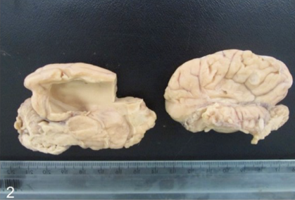
Pneumocephalus is a well described disease; it is commonly diagnosed in humans, but the condition is rarely encountered in veterinary medicine. Computed tomography (CT) is the gold-standard diagnostic method for identifying this disease, and other methods (such as necropsy) are rarely described. In this report, we describe necropsy findings of a 10-month-old, mixed-breed dog with intraventricular pneumocephalus. The dog was referred to Laboratory of Animal Pathology of the Federal University of Uberlândia, Brazil, for necropsy after being diagnosed with pneumocephalus upon CT. In the examination, the brain had dilation of both lateral ventricles with empty spaces. Histopathology showed congestion and mineralization only near the lateral ventricles, leading to the diagnosis of pneumocephalus based on the macroscopic findings. The animal also showed sinusitis characterized by nasal discharge and neutrophilic infiltration of nasal sinuses. However, bacterial culture was not conclusive because of contamination of the sample. This is therefore an important report that shows necropsy findings of intraventricular pneumocephalus, which is a rare condition in dogs. By documenting the necropsy findings, we hope to help veterinary pathologists, including those with limited access to diagnostic imaging.
Downloads
Downloads
Publicado
Edição
Seção
Licença
Copyright (c) 2022 Acta Veterinaria Brasilica

Este trabalho está licenciado sob uma licença Creative Commons Attribution 4.0 International License.
Autores que publicam na Acta Veterinaria Brasilica concordam com os seguintes termos: a) Autores mantém os direitos autorais e concedem à revista o direito de primeira publicação, com o trabalho simultaneamente licenciado sob a Licença Creative Commons Attribution que permite o compartilhamento do trabalho com reconhecimento da autoria e publicação inicial nesta revista. b) Autores têm autorização para assumir contratos adicionais separadamente, para distribuição não-exclusiva da versão do trabalho publicada nesta revista (ex.: publicar em repositório institucional ou como capítulo de livro), com reconhecimento de autoria e publicação inicial nesta revista. c) Autores têm permissão e são estimulados a publicar e distribuir seu trabalho online (ex.: em repositórios institucionais ou na sua página pessoal) a qualquer ponto antes ou durante o processo editorial, já que isso pode gerar alterações produtivas, bem como aumentar o impacto e a citação do trabalho publicado (Veja O Efeito do Acesso Livre).

 Esta obra está licenciada com uma Licença