
Holoprosencephaly associated with synophthalmia and arrhinia in feline (Felis catus): a case report
DOI:
https://doi.org/10.21708/avb.2023.17.3.11602Resumo
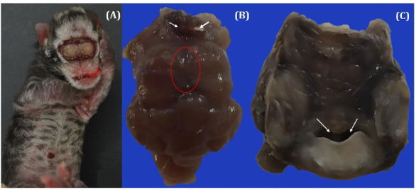
The aim of this study was to describe the macroscopic and histological lesions observed in domestic newborn felines with facial lesions characterized by holoprosencephaly associated with synophthalmia and arhinia. A feline with multiple craniofacial congenital anomalies was necropsied, and the brain was collected, followed by photodocumentation and histopathological processing with H&E. The diagnosis of alobar holoprosencephaly with grade (I) morphological lesions of the face was made through the anatomopathological changes observed during the clinical and necroscopic examination, characterized by deformation of the cerebral hemispheres, shown failure in the forebrain vesicle development, located in the cranial neural tube part, associated with craniofacial dysmorphisms. In the microscopic study of the CNS, lesions characterized by abnormal lamination of the cerebral hemisphere's outermost cell layers and multiple areas of vacuolization of the cortex and in the cerebellum low cell density of the granular layer, with the presence of large intracytoplasmic vacuoles and absence of Purkinje cells were observed. It was not possible to determine the deformation cause, since laboratory tests were not performed, as well as the association with the FeLV virus.
Downloads
Downloads
Publicado
Edição
Seção
Licença
Copyright (c) 2023 Acta Veterinaria Brasilica

Este trabalho está licenciado sob uma licença Creative Commons Attribution 4.0 International License.
Autores que publicam na Acta Veterinaria Brasilica concordam com os seguintes termos: a) Autores mantém os direitos autorais e concedem à revista o direito de primeira publicação, com o trabalho simultaneamente licenciado sob a Licença Creative Commons Attribution que permite o compartilhamento do trabalho com reconhecimento da autoria e publicação inicial nesta revista. b) Autores têm autorização para assumir contratos adicionais separadamente, para distribuição não-exclusiva da versão do trabalho publicada nesta revista (ex.: publicar em repositório institucional ou como capítulo de livro), com reconhecimento de autoria e publicação inicial nesta revista. c) Autores têm permissão e são estimulados a publicar e distribuir seu trabalho online (ex.: em repositórios institucionais ou na sua página pessoal) a qualquer ponto antes ou durante o processo editorial, já que isso pode gerar alterações produtivas, bem como aumentar o impacto e a citação do trabalho publicado (Veja O Efeito do Acesso Livre).

 Esta obra está licenciada com uma Licença