
Anatomy of the spinal cord of Alouatta belzebul
DOI:
https://doi.org/10.21708/avb.2018.12.2.7349Resumo
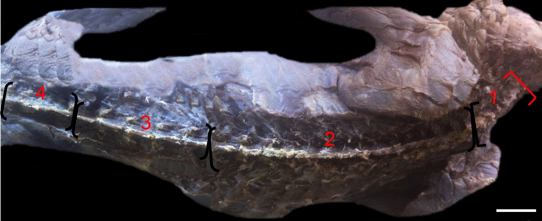
The genus Alouatta hosts species popularly known as red-handed howler, presenting wide geographic distribution and being found in several biomes. The objective was describe the anatomy of spinal cord of Alouatta belzebul specimens, focusing on the topography of medullary cone, stressing the cervical and lumbar intumescences and cauda equina, to provide anatomical data and compare it with other species to assist in anesthetic and surgical procedures. Four animals were received for scientific research, post mortem, from the fauna rescue program of Hydroelectric Plant of Belo Monte, Pará, and they were fixed in 10% formaldehyde solution. Structures such as the medullary cone, cervical and lumbar intumescence, and cauda equina were photographed (Sony α200-10.2 mpx). After thawing, we measured the specimens and observed a size of 80 to 82 cm from head to toe. After the skin and musculature were removed, it was observed that the spine of all specimens presented 7 cervical, 13 thoracic, 5 lumbar and 3 fused sacral vertebrae. The spinal cord was exposed after the removal of vertebral arches, it has 22 cm length in all animals, presenting the cervical intumescence between C3 and C6 vertebrae, with average of 2.2 cm and lumbar intumescence between T11 and T12 vertebrae, with average of 1.65 cm. The medullary cone is located between T12 and L1 vertebrae, with average of 1.5 cm, and the cauda equina between L1 and S3, with an average of 15 cm. This study has an important role as the basis for epidural anesthesia in the species.
Downloads
Downloads
Publicado
Edição
Seção
Licença
Autores que publicam na Acta Veterinaria Brasilica concordam com os seguintes termos: a) Autores mantém os direitos autorais e concedem à revista o direito de primeira publicação, com o trabalho simultaneamente licenciado sob a Licença Creative Commons Attribution que permite o compartilhamento do trabalho com reconhecimento da autoria e publicação inicial nesta revista. b) Autores têm autorização para assumir contratos adicionais separadamente, para distribuição não-exclusiva da versão do trabalho publicada nesta revista (ex.: publicar em repositório institucional ou como capítulo de livro), com reconhecimento de autoria e publicação inicial nesta revista. c) Autores têm permissão e são estimulados a publicar e distribuir seu trabalho online (ex.: em repositórios institucionais ou na sua página pessoal) a qualquer ponto antes ou durante o processo editorial, já que isso pode gerar alterações produtivas, bem como aumentar o impacto e a citação do trabalho publicado (Veja O Efeito do Acesso Livre).

 Esta obra está licenciada com uma Licença