
Feline sarcoid: case report
DOI:
https://doi.org/10.21708/avb.2020.14.3.8965Resumo
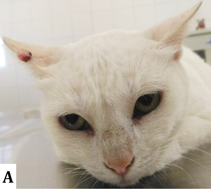
It was aimed to describe the clinical and histopathological characteristics of a case of sarcoid tumor in an adult, mixed-breed female cat, with a history of a small increase of volume on the ear. At physical examination, no alteration was found with the exception of the presence of a rounded dermal nodule of 3.0 x 1.5 x 0.5 cm, ulcerated, well-circumscribed, pedunculated, pinkish, solid-elastic, mobile and painless, located on the skin of the border of the auricular pavilion. In the cytological examination of the nodule, the involvement of a benign mesenchymal neoplasia was found. Furthermore, alterations in the hematological examinations were not observed, nor in the imaging tests, and the serological analyses for infection by the leukemia and immunodeficiency were negative. The surgical excision of the nodule was performed, and then the nodule was submitted to histopathologic examination. Microscopically, was verified superficial and deep dermis distended by a non-encapsulated nodule, covered by intact skin, constituted by fibroblasts disposed in random beams predominantly spaced, at times, interspersed by collagen fibers; epidermis with focally extensive acanthosis, projecting papillae in the direction of the superficial dermis. Furthermore, was observed accentuated orthokeratotic hyperkeratosis and, in occasional vessels, the presence of a discreet lymphoplasmocitary infiltrate and an area of focal hemorrhage, compatible with feline sarcoid. 11 months after the conchectomy, relapses have not been observed. This report points to the need of including this neoplasm in the differential diagnosis of cutaneous neoformations in cats with the aforementioned characteristics.
Downloads
Downloads
Publicado
Edição
Seção
Licença
Autores que publicam na Acta Veterinaria Brasilica concordam com os seguintes termos: a) Autores mantém os direitos autorais e concedem à revista o direito de primeira publicação, com o trabalho simultaneamente licenciado sob a Licença Creative Commons Attribution que permite o compartilhamento do trabalho com reconhecimento da autoria e publicação inicial nesta revista. b) Autores têm autorização para assumir contratos adicionais separadamente, para distribuição não-exclusiva da versão do trabalho publicada nesta revista (ex.: publicar em repositório institucional ou como capítulo de livro), com reconhecimento de autoria e publicação inicial nesta revista. c) Autores têm permissão e são estimulados a publicar e distribuir seu trabalho online (ex.: em repositórios institucionais ou na sua página pessoal) a qualquer ponto antes ou durante o processo editorial, já que isso pode gerar alterações produtivas, bem como aumentar o impacto e a citação do trabalho publicado (Veja O Efeito do Acesso Livre).

 Esta obra está licenciada com uma Licença