Esophageal diverticulum in a young feline: anatomopathological characterization
DOI:
https://doi.org/10.21708/avb.2025.19.2.12398Resumo
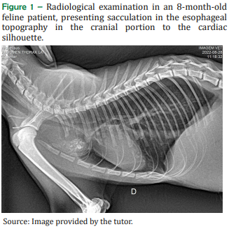
Esophageal diverticula are sac-like dilations in the esophageal wall, rare in felines, and classified as congenital or acquired. An 8-month-old male, mongrel cat presented with regurgitation, dysphagia, emesis, and reflux. Radiologic examination revealed a sacculation in the esophageal topography in the cranial portion of the cardiac silhouette. The patient underwent a gastrostomy to place a tube. Subsequently, he underwent esophageal endoscopic surgery, and diverticula of the thoracic esophagus and an underlying stenosis were identified. There was no resistance during the endoscopic passage between the esophagus and stomach, indicating a probable incompetence of the lower esophageal sphincter. The patient died post-surgery and was referred for necropsy. On macroscopic examination, the esophagus presented moderate dilatation in the medial portion, which, on cross-section, was formed by multiple diverticula. Caudal to the diverticula, an area of esophageal stenosis was noted. Microscopically, the esophageal diverticula had mucosa, submucosa, muscular and adventitia, and necro suppurative esophagitis. Thus, the present study aims to report a case of esophageal diverticulum associated with esophageal stenosis in a young feline, characterizing its anatomopathological aspects.
Downloads
Publicado
Edição
Seção
Licença
Copyright (c) 2025 Acta Veterinaria Brasilica

Este trabalho está licenciado sob uma licença Creative Commons Attribution 4.0 International License.
Autores que publicam na Acta Veterinaria Brasilica concordam com os seguintes termos: a) Autores mantém os direitos autorais e concedem à revista o direito de primeira publicação, com o trabalho simultaneamente licenciado sob a Licença Creative Commons Attribution que permite o compartilhamento do trabalho com reconhecimento da autoria e publicação inicial nesta revista. b) Autores têm autorização para assumir contratos adicionais separadamente, para distribuição não-exclusiva da versão do trabalho publicada nesta revista (ex.: publicar em repositório institucional ou como capítulo de livro), com reconhecimento de autoria e publicação inicial nesta revista. c) Autores têm permissão e são estimulados a publicar e distribuir seu trabalho online (ex.: em repositórios institucionais ou na sua página pessoal) a qualquer ponto antes ou durante o processo editorial, já que isso pode gerar alterações produtivas, bem como aumentar o impacto e a citação do trabalho publicado (Veja O Efeito do Acesso Livre).

 Esta obra está licenciada com uma Licença