Evaluation of the Ki‐67 cell proliferation index as a prognostic factor in female dogs with simple mammary carcinomas of papillary and tubular subtypes
DOI:
https://doi.org/10.21708/avb.2025.19.3.12926Abstract
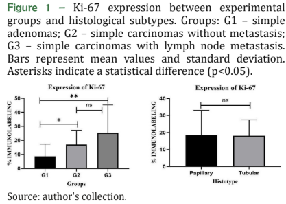
Mammary tumors are the most prevalent neoplasms in female dogs, representing a significant clinical issue. Ki‐67 is the most studied biomarker for tumor proliferation and is related to the patient’s prognosis in many tumor types. This study aimed to determine the most efficient cutoff point of the immunohistochemical marker Ki‐67 to establish the prognosis of female dogs with simple mammary papillary and tubular carcinomas. Additionally, to compare their expressions in different clinicopathological situations. Mammary tissue samples were divided into three experimental groups: G1 (simple adenomas, n = 19), G2 (simple carcinomas without metastasis, n = 43), and G3 (simple carcinomas with metastasis in lymph nodes, n = 8). The samples were subjected to immunohistochemistry using the MIB‐1 antibody (Dako®) to compare Ki‐67 expression, histological subtypes, and clinicopathological variables. Subsequently, different Ki‐67 cutoff points were analyzed in regard to overall patient survival. Simple carcinomas showed more intense Ki‐67 labeling than simple adenomas (P<0.05). Ki‐67 expression below 24% did not correlate with overall survival. However, an expression level of 27% (p = 0.0048) and 33% (p = 0.0025) significantly correlated with a shorter overall survival time, and 100% of the dogs with a Ki‐67 expression greater than 33% were deceased at the end of the follow‐up period. Higher percentages of Ki‐67 labeling were observed in high‐grade tumors, those larger than 3 centimeters and with a high mitotic index. Therefore, a Ki‐67 above 27% can be used to indicate the prognosis and help in therapeutic decision‐making in female dogs with simple mammary carcinomas.
Dowloads
Pubblicato
Fascicolo
Sezione
Licenza
Copyright (c) 2026 Acta Veterinaria Brasilica

Questo volume è pubblicato con la licenza Creative Commons Attribuzione 4.0 Internazionale.
Autores que publicam na Acta Veterinaria Brasilica concordam com os seguintes termos: a) Autores mantém os direitos autorais e concedem à revista o direito de primeira publicação, com o trabalho simultaneamente licenciado sob a Licença Creative Commons Attribution que permite o compartilhamento do trabalho com reconhecimento da autoria e publicação inicial nesta revista. b) Autores têm autorização para assumir contratos adicionais separadamente, para distribuição não-exclusiva da versão do trabalho publicada nesta revista (ex.: publicar em repositório institucional ou como capítulo de livro), com reconhecimento de autoria e publicação inicial nesta revista. c) Autores têm permissão e são estimulados a publicar e distribuir seu trabalho online (ex.: em repositórios institucionais ou na sua página pessoal) a qualquer ponto antes ou durante o processo editorial, já que isso pode gerar alterações produtivas, bem como aumentar o impacto e a citação do trabalho publicado (Veja O Efeito do Acesso Livre).

 Esta obra está licenciada com uma Licença