
Macro, micromorphological and histological aspects of the intestine pirarucu Arapaima gigas (SCHINZ, 1822) (Osteoglossiformes: Arapaimidae)
DOI:
https://doi.org/10.21708/avb.2022.16.4.10728Resumo
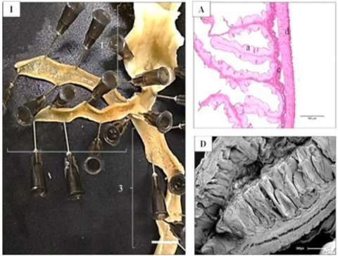
Currently, the fish farming with Arapaima gigas has suffered from technological obstacles in the fields of reproduction, health and nutrition, making it necessary to know the morphology of its structures, so that it can advance in more technified research in scope of production in Rondônia state, as well as in aquaculture nutrition and health. Therefore, the aimed is to characterize the macro and microscopic morphology of posterior digestive system of A. gigas. The intestine of six specimens A. gigas in ideal slaughter size was analyzed. The analyzes were performed using light-sheet microscopy (LM) and scanning electron (SEM) techniques. The intestine basically showes similar histological characteristics in three analyzed portions (proximal, middle and distal). Same type of simple columnar epithelium with goblet cells was evidenced, with subtle variations in pattern of villi in each segment, and in number of goblet cells. In the rectum, the amount of goblet cells and evident longitudinal villi was expressive. Macroscopic anatomy and histology of the intestine A. gigas analyzed showes characteristics of adaptation to cultivation, according to their diet and habitat. The intestinal mucosa can divided into three distinct portions: proximal, middle and final intestine, in addition to the rectum and anus. In the pyloric cecum, the folds are slightly higher and poorly branched. The rectum, compared to the midgut, showed a higher occurrence of goblet cells in the mucosa. This increase in goblet cells observed in the posterior portion may related to the assimilation of ions and fluids that occur at this location.
Downloads
Downloads
Publicado
Edição
Seção
Licença
Copyright (c) 2022 Acta Veterinaria Brasilica

Este trabalho está licenciado sob uma licença Creative Commons Attribution 4.0 International License.
Autores que publicam na Acta Veterinaria Brasilica concordam com os seguintes termos: a) Autores mantém os direitos autorais e concedem à revista o direito de primeira publicação, com o trabalho simultaneamente licenciado sob a Licença Creative Commons Attribution que permite o compartilhamento do trabalho com reconhecimento da autoria e publicação inicial nesta revista. b) Autores têm autorização para assumir contratos adicionais separadamente, para distribuição não-exclusiva da versão do trabalho publicada nesta revista (ex.: publicar em repositório institucional ou como capítulo de livro), com reconhecimento de autoria e publicação inicial nesta revista. c) Autores têm permissão e são estimulados a publicar e distribuir seu trabalho online (ex.: em repositórios institucionais ou na sua página pessoal) a qualquer ponto antes ou durante o processo editorial, já que isso pode gerar alterações produtivas, bem como aumentar o impacto e a citação do trabalho publicado (Veja O Efeito do Acesso Livre).

 Esta obra está licenciada com uma Licença