
Using 3D computed tomography in the anatomical description of the eye and the vestibulocochlear organ of a blue-and-yellow macaw (Ara ararauna Linnaeus, 1758) and of a toucan (Ramphastos toco Statius Muller, 1776)
DOI:
https://doi.org/10.21708/avb.2020.14.4.9464Resumo
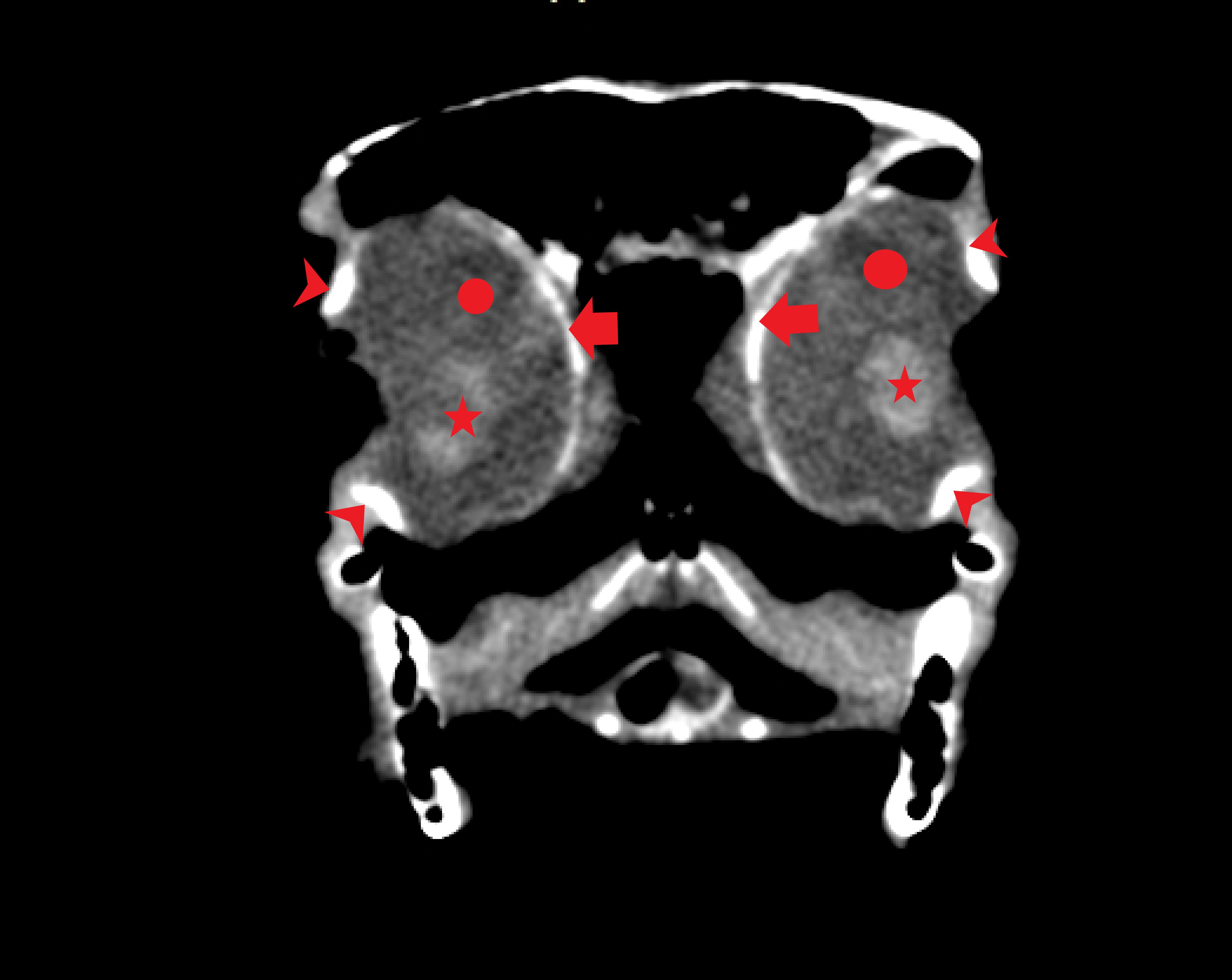
The aim of this study was to evaluate the use of Computed Tomography to study the anatomy of the eye and the vestibulocochlear organ of the wild birds. For this purpose, formaldehyde-embalmed specimens of a toucan and of a blue-and-yellow macaw were submitted to a whole-body scan by a 64 slice-Multidetector CT yielding 0,7mm-thick transversally oriented images. These were reconstructed by specific software that produced additional images in dorsal, transversal, and sagittal planes, as well as three-dimensional images, which were obtained by two techniques: Maximum Intensity Projection and Volume Rendering. Our study found that the eye bulbs in the orbit occupy a proportionally large space in the skull, highlighting the important role that vision plays in these animals. CT provided gross anatomic information about the size and shape of the eye, such as lenses and scleral rings of these birds. Regarding the vestibulocochlear organ, CT was less likely to identify the inner ear structures, especially the ones of the membranous labyrinth. The bony semicircular canals were clearly seen and in the middle ear, the columella was identified. Our results demonstrate that the vestibulocochlear organ of birds is less complex than that of mammals, although, as expected, the semicircular canals are very well developed, being adapted to the accurate balance present in these animals. CT can be used as a good technique to evaluate eye and ear structures on these birds, and can be useful to study them in vivo for pathological conditions or for comparisons between different species.
Downloads
Downloads
Publicado
Edição
Seção
Licença
Autores que publicam na Acta Veterinaria Brasilica concordam com os seguintes termos: a) Autores mantém os direitos autorais e concedem à revista o direito de primeira publicação, com o trabalho simultaneamente licenciado sob a Licença Creative Commons Attribution que permite o compartilhamento do trabalho com reconhecimento da autoria e publicação inicial nesta revista. b) Autores têm autorização para assumir contratos adicionais separadamente, para distribuição não-exclusiva da versão do trabalho publicada nesta revista (ex.: publicar em repositório institucional ou como capítulo de livro), com reconhecimento de autoria e publicação inicial nesta revista. c) Autores têm permissão e são estimulados a publicar e distribuir seu trabalho online (ex.: em repositórios institucionais ou na sua página pessoal) a qualquer ponto antes ou durante o processo editorial, já que isso pode gerar alterações produtivas, bem como aumentar o impacto e a citação do trabalho publicado (Veja O Efeito do Acesso Livre).

 Esta obra está licenciada com uma Licença