
Mucosa cytopathology for research of Sinegaglia-Lentz corpuscles in canine distemper
DOI:
https://doi.org/10.21708/avb.2020.14.2.9315Resumo
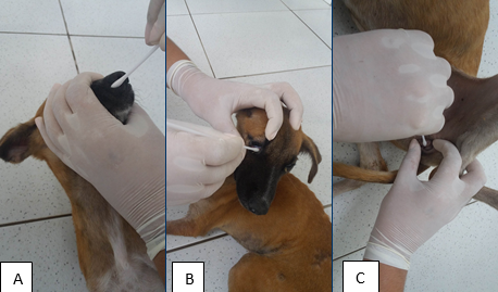
Canine distemper (CD) is a multisystemic and infectious disease caused by a Morbillivirus. The search for viral inclusions by mucosal cytology is a low cost and high practical alternative, which establishes the definitive diagnosis of CD, but is little reported in the literature. The objective was to encourage the use of mucosal cytopathology to identify Sinegaglia-Lentz corpuscles in the veterinary routine as an alternative or complementary to blood screening, providing a selection of photographs of the viral inclusions. Selected were 16 dogs with classic systemic or neurological disorders and positive for the chromatographic immunoassay for CD. Samples of the conjunctival, nasal and genital epithelium were collected with the aid of a sterile swab for making slides. Whole blood was also collected to make a blood smear. The slides were stained with a fast panoptic and observed by optical microscopy to directly search for eosinophilic viral inclusions, at 40 and 100x magnification. Sinegaglia-Lentz corpuscles were detected in nine of the 16 dogs (56.25%), five in conjunctival mucosa (41.65%), three in nasal mucosa (25%), one in genital mucosa (8.33%) and three in blood smear (25%). It is concluded that mucosa cytopathology, especially conjunctival, for Sinegaglia-Lentz research is an auxiliary tool for the early and definitive diagnosis of canine distemper. However, the absence of viral inclusions in these samples does not rule out the possibility of the disease.
Downloads
Downloads
Publicado
Edição
Seção
Licença
Autores que publicam na Acta Veterinaria Brasilica concordam com os seguintes termos: a) Autores mantém os direitos autorais e concedem à revista o direito de primeira publicação, com o trabalho simultaneamente licenciado sob a Licença Creative Commons Attribution que permite o compartilhamento do trabalho com reconhecimento da autoria e publicação inicial nesta revista. b) Autores têm autorização para assumir contratos adicionais separadamente, para distribuição não-exclusiva da versão do trabalho publicada nesta revista (ex.: publicar em repositório institucional ou como capítulo de livro), com reconhecimento de autoria e publicação inicial nesta revista. c) Autores têm permissão e são estimulados a publicar e distribuir seu trabalho online (ex.: em repositórios institucionais ou na sua página pessoal) a qualquer ponto antes ou durante o processo editorial, já que isso pode gerar alterações produtivas, bem como aumentar o impacto e a citação do trabalho publicado (Veja O Efeito do Acesso Livre).

 Esta obra está licenciada com uma Licença