Clinical, surgical, and radiographic aspects in a white-eared opossum (Didelphis albiventris) with traumatic diaphragmatic hernia
DOI:
https://doi.org/10.21708/avb.2021.15.4.10259Abstract
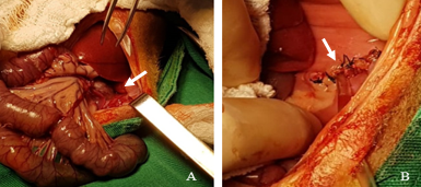
This is the case of a specimen of Didelphis albiventris with signs of respiratory difficulty after a dog attack. Thoracic radiographic examination revealed pneumothorax, pulmonary contusion, and rib fracture, but no alteration compatible with diaphragmatic hernia was observed. Pneumothorax was reduced and the other alterations were treated. However, clinical manifestations persisted, and thus a contrast-gastrointestinal radiographic study was performed, showing abdominal organs in the thoracic cavity and loss of diaphragmatic line. The surgical approach was instituted, with access to the diaphragm through median laparotomy. Through the diaphragmatic rupture, present in the left antimere, there were herniated liver and gastric portions, intestinal segments, and omentum. After inspection and repositioning of the abdominal organs, the diaphragm raffia was performed with single sutures interrupted with 3-0 Nylon thread. The patient's complete recovery occurred 14 days after the surgical procedure, with remission of clinical manifestations and normality of thoracic images in radiographic studies.
Downloads
Downloads
Published
Issue
Section
License
Autores que publicam na Acta Veterinaria Brasilica concordam com os seguintes termos: a) Autores mantém os direitos autorais e concedem à revista o direito de primeira publicação, com o trabalho simultaneamente licenciado sob a Licença Creative Commons Attribution que permite o compartilhamento do trabalho com reconhecimento da autoria e publicação inicial nesta revista. b) Autores têm autorização para assumir contratos adicionais separadamente, para distribuição não-exclusiva da versão do trabalho publicada nesta revista (ex.: publicar em repositório institucional ou como capítulo de livro), com reconhecimento de autoria e publicação inicial nesta revista. c) Autores têm permissão e são estimulados a publicar e distribuir seu trabalho online (ex.: em repositórios institucionais ou na sua página pessoal) a qualquer ponto antes ou durante o processo editorial, já que isso pode gerar alterações produtivas, bem como aumentar o impacto e a citação do trabalho publicado (Veja O Efeito do Acesso Livre).
 Esta obra está licenciada com uma Licença