
Disseminated histoplasmosis in an urban canine with access to the rural environment
DOI:
https://doi.org/10.21708/avb.2024.18.1.11852Abstract
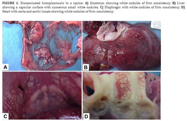
Histoplasmosis is a systemic disease caused by the fungus Histoplasma capsulatum. The microorganism causes localized granulomatous disease, with the respiratory tract being most affected, and can spread through the lymphatic and blood stream. Severe infection in dogs is associated with immunosuppressive therapies. The reported case occurred in a canine, brought to care with a serious clinical condition, which did not respond to treatment and was euthanized. The necropsy examination showed white nodules, of firm consistency and distributed throughout the omentum, liver, splenic capsule, pancreas, stomach serosa, intestines, bladder, mesenteric and inguinal lymph nodes, diaphragm, parietal pleura, aorta and aortic lunate. Microscopically, all nodular lesions consisted of areas of necrosis, with intense histiolymphoplasmacytic infiltrate, epithelioid macrophages and Langhans-type giant cells with yeast-like structures, with morphology and arrangement consistent with Histoplasma spp., confirmed by special Periodic Acid-Schiff (PAS) stains and Grocott. Therefore, the present study aims to describe a case of multisystemic histoplasmosis in a canine, diagnosed through anatomopathology and histochemistry, and to alert to this potential infection in urban canines with access to rural areas and/or environments with the fungal agent.
Downloads
Downloads
Published
Issue
Section
License
Copyright (c) 2024 Acta Veterinaria Brasilica

This work is licensed under a Creative Commons Attribution 4.0 International License.
Autores que publicam na Acta Veterinaria Brasilica concordam com os seguintes termos: a) Autores mantém os direitos autorais e concedem à revista o direito de primeira publicação, com o trabalho simultaneamente licenciado sob a Licença Creative Commons Attribution que permite o compartilhamento do trabalho com reconhecimento da autoria e publicação inicial nesta revista. b) Autores têm autorização para assumir contratos adicionais separadamente, para distribuição não-exclusiva da versão do trabalho publicada nesta revista (ex.: publicar em repositório institucional ou como capítulo de livro), com reconhecimento de autoria e publicação inicial nesta revista. c) Autores têm permissão e são estimulados a publicar e distribuir seu trabalho online (ex.: em repositórios institucionais ou na sua página pessoal) a qualquer ponto antes ou durante o processo editorial, já que isso pode gerar alterações produtivas, bem como aumentar o impacto e a citação do trabalho publicado (Veja O Efeito do Acesso Livre).

 Esta obra está licenciada com uma Licença