
Ultrasound methods used for planning for cataract surgery in dogs
DOI:
https://doi.org/10.21708/avb.2019.13.2.8332Abstract
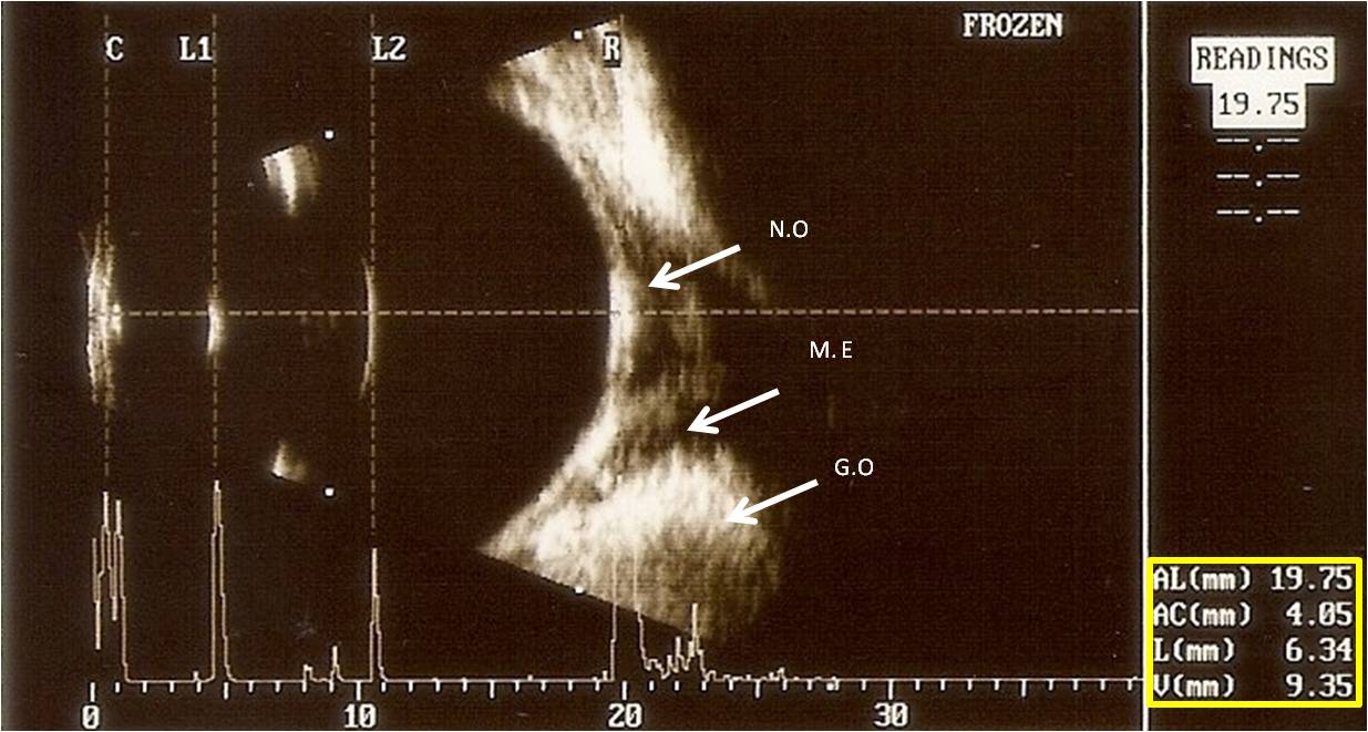
Cataract is an important disease on veterinary ophthalmology. Different techniques facilitate the classification of the cataracts, adding information obtained by the conventional eye exam. This review discusses different modalities of ultrasonography methods available for diagnosis and surgical planning in dogs with cataracts. A-Mode ultrasound is used to evaluate thickness and length of the lens. B-Mode also has the capacity to evaluate features of echogenicity, echotexture, position of the lens and the identification of retinal and vitreous degenerations. The ultrasonic biomicroscopy is useful in assessing the anterior capsule and the positioning of the lens. The Doppler method it is useful to detection of vascular changes in patients with cataracts. The elastography method however allows measuring the lens rigidity, which is extremely important for planning and surgical prognosis.
Downloads
Downloads
Published
Issue
Section
License
Autores que publicam na Acta Veterinaria Brasilica concordam com os seguintes termos: a) Autores mantém os direitos autorais e concedem à revista o direito de primeira publicação, com o trabalho simultaneamente licenciado sob a Licença Creative Commons Attribution que permite o compartilhamento do trabalho com reconhecimento da autoria e publicação inicial nesta revista. b) Autores têm autorização para assumir contratos adicionais separadamente, para distribuição não-exclusiva da versão do trabalho publicada nesta revista (ex.: publicar em repositório institucional ou como capítulo de livro), com reconhecimento de autoria e publicação inicial nesta revista. c) Autores têm permissão e são estimulados a publicar e distribuir seu trabalho online (ex.: em repositórios institucionais ou na sua página pessoal) a qualquer ponto antes ou durante o processo editorial, já que isso pode gerar alterações produtivas, bem como aumentar o impacto e a citação do trabalho publicado (Veja O Efeito do Acesso Livre).

 Esta obra está licenciada com uma Licença