Clinical and anatomopathological diagnosis in bovines affected by perforated abomasal ulcers and with comorbities
DOI:
https://doi.org/10.21708/avb.2023.17.3.11731Resumen
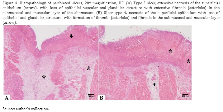
This study describes the findings of the clinical and anatomopathological diagnosis in cattle affected by perforated abomasal ulcers and with comorbidities. Twenty-five cattle were evaluated, young (n=8, aged less than two years) and adults (n=17, aged greater than two and a half years), diagnosed with perforated abomasal ulcers. Epidemiological information was retrieved from the medical records. Blood, ruminal fluid, and peritoneal fluid (PI) samples were collected for laboratory examination. Ultrasound examination, exploratory laparotomy, and post mortem examinations were performed for diagnostic purposes. The main clinical findings of perforated ulcers in the present study were: apathy, dehydration, intestinal hypomotility, diarrheal stools, hypomotile or atonic rumen, tachycardia, tachypnea, abdominal distention with bilateral bulging, and increased abdominal tension. The most frequent abnormal hematological findings were hypoproteinemia and neutrophilic leukocytosis with a left shift. The peritoneal fluid (PL) showed an inflammatory reaction in the cytology, characteristic of an exudate. A confirmatory diagnosis of peritonitis by ultrasonography was possible in five cases, and in two cases at exploratory laparotomy. In the post mortem examination, the ulcers were characterized as type 3, 4, or 5, with comorbidities present in 68% of the cases. It is necessary to combine ultrasonography with abdominal centesis and exploratory laparotomy procedures to suggest perforated ulcers. However, post mortem examination better characterizes the type of perforated ulcer present, particularly type 5.
Descargas

Descargas
Publicado
Número
Sección
Licencia
Derechos de autor 2023 Acta Veterinaria Brasilica

Esta obra está bajo una licencia internacional Creative Commons Atribución 4.0.
Autores que publicam na Acta Veterinaria Brasilica concordam com os seguintes termos: a) Autores mantém os direitos autorais e concedem à revista o direito de primeira publicação, com o trabalho simultaneamente licenciado sob a Licença Creative Commons Attribution que permite o compartilhamento do trabalho com reconhecimento da autoria e publicação inicial nesta revista. b) Autores têm autorização para assumir contratos adicionais separadamente, para distribuição não-exclusiva da versão do trabalho publicada nesta revista (ex.: publicar em repositório institucional ou como capítulo de livro), com reconhecimento de autoria e publicação inicial nesta revista. c) Autores têm permissão e são estimulados a publicar e distribuir seu trabalho online (ex.: em repositórios institucionais ou na sua página pessoal) a qualquer ponto antes ou durante o processo editorial, já que isso pode gerar alterações produtivas, bem como aumentar o impacto e a citação do trabalho publicado (Veja O Efeito do Acesso Livre).


 Esta obra está licenciada com uma Licença